Ricardo Insausti Neuroanatomical Archive
Ricardo Insausti (1954-2024)
Ricardo Insausti was among the most influential neuroanatomists of his generation. Over a career that spanned five decades, he studied and extensively described the cytoarchitecture, organization, and connectivity of the medial temporal lobe in humans, non-human primates, and rodents. His work profoundly shaped our understanding of the structure and function of this critical brain region, which plays a central role in memory and is of key relevance in neurodegenerative disease research. At the head of the Human Neuroanatomy Laboratory (HNL) at the University of Castilla La Mancha, Ricardo oversaw a major brain bank and established novel techniques for in situ brain perfusion and tissue preparation optimized for neuroanatomical studies. He was a key contributor and strong supporter of the effort by the Hippocampal Subfields Group (HSG) to develop a harmonized, reliable protocol for medial temporal lobe segmentation on in vivo MRI.
Ricardo was a prolific author, educator, and collaborator who taught multiple generations of future neuroscientists and physicians. His trainees and colleagues fondly remember his kindness, uncommon generosity and a genuine passion for science. Ricardo believed that science should be open to all and unimpeded by boundaries. In that spirit, this archive provides open access to a large library of neuroanatomical annotations created by Ricardo and his colleagues, preserving his legacy and supporting future generations of researchers.

The Neuroanatomical Archive
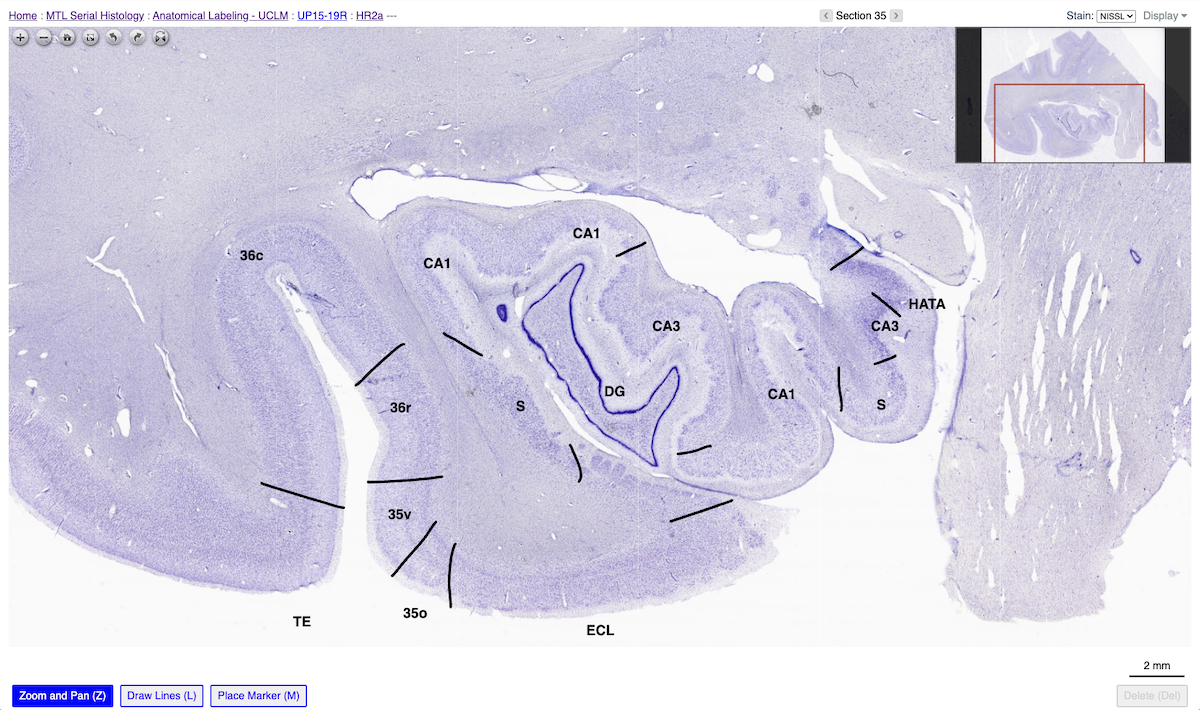
Between 2019 and 2024, Ricardo led a team of neuroanatomists at HNL in generating fine-grained annotations of medial temporal lobe subregions across a large collection of digital histology images, serially sectioned along the length of the temporal lobe, creating an unparalleled resource of neuroanatomical knowledge. This work was carried out as part of a collaboration with researchers at the Alzheimer’s Disease Research Center at the University of Pennsylvania on an NIH-funded project (R01 AG056014) aiming to characterize the anatomical variability and spread of neurodegenerative pathologies in the medial temporal lobe using matching postmortem MRI and serial histology. This archive makes these annotations openly available to the neuroscience research community.
The archive contains complete long-axis annotations of the medial temporal lobe for 65 specimens, with >124,000 annotations across >8,500 slides. Histological sections were obtained from brain donations at the University of Castilla-La Mancha and the University of Pennsylvania. Consent from the next of kin was obtained in all brain donations. The intact temporal lobe was sectioned serially using a custom 3D printed mold derived from postmortem MRI, with 50µm thick Nissl-stained sections obtained every 500µm and digitally scanned at 20x resolution (Insausti et al., 2023). Ricardo and his colleagues at HNL annotated the boundaries of >25 fine-grained MTL subregions. These annotations were used to generate 3D probabilistic atlases of medial temporal lobe anatomy and quantify tau pathology across MTL subregions (Ravikumar et al., 2024) and are being leveraged by the Hippocampal Subfields Group to inform the development of a harmonized protocol for MTL segmentation on in vivo MRI.
Bibliography and Additional Resources
-
Lorduy, M.C., Insausti Serrano, A.M., Muñoz López, M., Wisse, L., Yushkevich, P.A. and Amaral, D.G. (2025) ‘Remembering Ricardo Serrano Insausti 1954–2024’, Hippocampus, 35(2), e70005. https://doi.org/10.1002/hipo.70005
-
Marcos Rabal, P., Arroyo Jiménez, M., Insausti Serrano, A.M., and Fornai, F. ‘In Memoriam of Prof. Dr. Ricardo Insausti (Pamplona, 1954–Albacete, 2024)’, Anatomia. https://www.mdpi.com/about/announcements/10673
-
Insausti, R. et al. (2023) ‘Ex vivo, in situ perfusion protocol for human brain fixation compatible with microscopy, MRI techniques, and anatomical studies’, Frontiers in Neuroanatomy, 17, Article 1149674. https://doi.org/10.3389/fnana.2023.1149674
-
Insausti, R. and Amaral, D.G. (2012) ‘Hippocampal formation’, in Mai, J.K. and Paxinos, G. (eds.) The Human Nervous System. 3rd edn. London: Academic Press, pp. 896–942. https://doi.org/10.1016/B978-0-12-374236-0.10024-0
-
Ravikumar, S. et al. (2024) ‘Postmortem imaging reveals patterns of medial temporal lobe vulnerability to tau pathology in Alzheimer’s disease’, Nature Communications, 15(1), Article 4803. https://doi.org/10.1038/s41467-024-49205-0
-
Wuestefeld, A. et al. (2024) ‘Comparison of histological delineations of medial temporal lobe cortices by four independent neuroanatomy laboratories’, Hippocampus, 34(5), pp. 241–260. https://doi.org/10.1002/hipo.23602
Software and Support
This archive uses the open-source PICSL Histology Annotation System (PHAS) platform. It was created and is maintained by Paul Yushkevich (UPenn). To report technical issues, please use the software’s issue tracker.